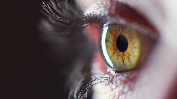
Researchers in a laboratory at the Faculty of Applied Sciences at Lausanne University EPFL have developed an eye medicine device that can be used to diagnose some degenerative eye disorders long before the first symptoms appear. According to the Neuroscience News website, which cited Ophthalmology Science, the initial model, in early clinical trials, demonstrated that the gadget generates a significant degree of accuracy in under five seconds.
Treatments to halt or slow the progression of degenerative eye diseases, which can result in blindness, are the subject of active research. However, there is currently no tool that can accurately identify these disorders before the onset of symptoms.
One of the most well-known degenerative eye conditions, AMD’s macro-age degeneration alters the eye’s optical receptors.
The degeneration of RPE, a layer of cells situated beneath optical receptors, is the fundamental cause of all degenerative eye diseases.
The new device, which was developed in the LAPD Applies Laboratory Laboratory, monitors the changes in the RPE retina epithelium before the symptoms appear, providing researchers the first images ever in the living body through which the cells can distinguish between cells.
Doctors will be able to diagnose these problems before the irreversible signs manifest themselves since the photos acquired by the novel equipment allow for early detection.
The researchers from EPFL created a network camera with two inclined beams that are trained on white light and an adaptive visual system that corrects for light wave aberrations to create a crisp image.
Transclerral optical imaging is a method that is similar to the infrared imaging systems now in use to take pictures of the retina.
But according to Professor Christophe Mosser, who heads the LAPD Applied Persons Laboratory at the University of Lausanne University, there is one major difference: “The packages are indirectly concentrated through the whites of the eye, which overcomes the problem of excessive light caused by the conical cells that are very reflected for light receptors When the retina is illuminated by the pupil, it is situated at the centre of the eye.
Then, in less than five seconds, the camera, which exits the eye through the pupil, captures the light waves. This is a fundamental speed feature for possible diagnostic application, and the camera can catch 100 raw photographs. The first photos are then collected and aligned by the algorithms to create a single, high-quality image on the screen.
A novel tool for the early detection of degenerative eye diseases

